Tissue-clearing protocols reduce optical scattering so that labeled structures can be imaged through thick tissue or intact organs. The central commissioning decision is chemical: the protocol must match the specimen, label, desired geometry, imaging medium, and downstream assay. This page compares clearing chemistry and specimen preservation. Light-sheet optics and whole-brain informatics are addressed separately.[S9][S11]
Why clear intact tissue
Physical sectioning samples selected planes and can interrupt continuous structures. CLARITY and iDISCO were developed to combine molecular labeling with a global three-dimensional view of intact tissue or large specimens.[S1][S3] Clearing is useful when continuity or spatial distribution is part of the endpoint. It does not replace section histology when cellular morphology, validated lesion grading, or routine pathology interpretation is the primary question.
A clearing workflow fixes and chemically transforms the specimen, adds or preserves contrast, matches refractive index, and then acquires a three-dimensional image. Reviews describe pairing cleared tissue with light-sheet microscopy and automated image analysis.[S9] Because clearing alters the specimen, “intact” means unsectioned, not chemically unchanged.
Clearing families and their tradeoffs
CUBIC
CUBIC uses immersion in chemical mixtures containing aminoalcohols and combines clearing with computational analysis. The original report demonstrated whole-brain imaging at single-cell resolution and described scalability from primate brain samples to subcellular structures.[S2] The source does not establish that every CUBIC implementation is highly reproducible, so reproducibility should be demonstrated for the laboratory, specimen, label, and analysis pipeline being commissioned.
CLARITY and DISCO-family methods
CLARITY converts fixed tissue into a nanoporous hydrogel hybrid, removes lipids, and permits antibody labeling through intact adult mouse brain; the original study also demonstrated repeated staining and destaining.[S1] iDISCO combines whole-mount immunolabeling with volume imaging of large cleared specimens and was reported compatible with 28 antibodies in the founding study.[S3] DISCO-family methods are organic-solvent based, as established for 3DISCO.[S4]
Protocol choice changes more than transparency. uDISCO was designed to preserve fluorescent proteins while shrinking intact organs and rodent bodies by as much as 65%.[S5] Other aqueous approaches emphasize fluorescence or geometry: Sca/e reported preservation of fluorescent signals,[S7] and SeeDB reported morphology-preserving clearing without quenching many fluorescent dyes.[S8] Compare dimensions before and after clearing whenever absolute distance, density, or volume is an endpoint.
Compatibility with imaging
The clearing and imaging plans must be designed together. Refractive index, chamber material, working distance, objective correction, specimen size, and mounting determine whether the cleared sample can actually be imaged. Tissue-clearing reviews describe light-sheet fluorescence microscopy as a common partner for intact-volume acquisition.[S9]
Preserving the signal that matters
Whole-brain molecular labeling
The original CUBIC report combined chemical clearing with whole-brain imaging and computational analysis at single-cell resolution and described scaling to primate brain tissue.[S2] That establishes technical feasibility under the reported conditions. A commissioned assay still needs tissue-specific acceptance criteria for transparency, label penetration, background, deformation, and analyzable depth.
For an immunolabeled endpoint, run a pilot that tests positive and negative controls, antibody penetration through the full depth, nonspecific background, lot effects, and whether signal intensity remains quantitative after clearing. iDISCO demonstrates broad antibody compatibility, but compatibility with a published panel does not validate a new antibody or tissue automatically.[S3]
Fluorescence, antigenicity, and morphology
If the endpoint depends on endogenous fluorescence, antigenicity, RNA, or fine tissue architecture, choose preservation chemistry explicitly. SHIELD uses polyfunctional crosslinking and was reported to preserve protein fluorescence, antigenicity, transcripts, and tissue architecture under harsh processing conditions.[S6] Sca/e emphasizes fluorescent-signal preservation,[S7] while SeeDB emphasizes morphology-preserving aqueous clearing.[S8] These are distinct design objectives, not interchangeable claims of overall superiority.
Commissioning the clearing workflow
Specify fixation, delipidation or decolorization, label, penetration control, refractive-index matching, dimensional calibration, mounting, image acceptance criteria, and raw-data delivery before the pilot begins. Protocol repositories note that adapting a clearing method to a new problem can require trial and error and that methods trade speed and cost against tissue stability and fluorescence quenching.[S11] For exploratory pharmacology or proof-of-concept work, FDA CBER guidance states that compliance with Good Laboratory Practice is not required for in vitro and in vivo pharmacology or POC studies.[S10] That statement concerns the individual study purpose; it is not a certification of a method or facility. If the images will support a regulated safety conclusion, define the applicable quality system and validation plan for that study.
Whole-brain imaging with single-cell resolution using chemical cocktails and computational analysis (CUBIC). https://europepmc.org/article/MED/24746791 (accessed 2026-09-03)
3 Recommended Contract Research Organizations
for Non-Clinical Studies
— by Target goal and Expertise
In non-clinical development, the choice of contract research organization shapes the quality of the data and the time it takes to reach the next decision. Below, three CROs are introduced by the type of study they support: pharmacology (efficacy) studies, safety studies, and pharmacokinetic (PK/PD) studies. Each summary describes the services the company offers so that you can match a provider to your target and development objective.
Pharmacology (Efficacy) StudiesDisease-Relevant Models for Translational Drug Evaluation
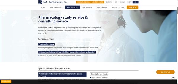
Reference: SMC Laboratories, Inc. official website (https://www.smccro-lab.com/)
SMC Laboratories, Inc.
SMC Laboratories is a specialized non-clinical CRO focused on in vivo pharmacology and efficacy studies using disease-relevant animal models, particularly in fibrosis, inflammation, metabolic diseases, and oncology.
Areas of Expertise
Disease-Relevant Model Portfolio
SMC Laboratories offers models covering the liver, lung, kidney, intestine, and oncology. Its portfolio includes the proprietary STAM™ model for MASH, fibrosis, and hepatocellular carcinoma.
Study Design Based on Target Biology
Study plans are developed around the target biology, mechanism of action, disease stage, and development objective. Pharmacological endpoints can be combined with histopathology, biomarkers, and disease-specific readouts.
Support from Target Validation to Proof of Concept
With experience from more than 1,000 studies for clients in 30 countries, SMC Laboratories supports programs from target validation and candidate selection through in vivo proof-of-concept studies.
Safety StudiesComprehensive Safety Assessment for Preclinical Development
Reference: Charles River Laboratories official website (https://www.criver.com/)
Charles River Laboratories
Charles River provides non-clinical toxicology and safety assessment services for programs ranging from exploratory safety studies to IND-enabling development.
Areas of Expertise
General Toxicology Across Study Designs
Services include single- and repeat-dose toxicology, dose-range finding, and general toxicology studies across multiple species and administration routes.
Non-GLP and GLP Study Support
Charles River supports both non-GLP and GLP studies, allowing sponsors to progress from early safety characterization to studies intended for regulatory submissions.
Integrated IND-Enabling Safety Assessment
Toxicology studies can be integrated with toxicokinetics, clinical pathology, histopathology, and safety pharmacology to support interpretation and IND-enabling safety packages.
Pharmacokinetic (PK/PD) StudiesConnecting Drug Exposure with Pharmacological Response
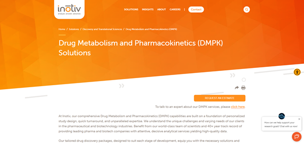
Reference: Inotiv official website (https://www.inotiv.com/)
Inotiv
Inotiv provides integrated PK/PD, DMPK, and bioanalytical services to characterize drug exposure and its relationship with pharmacological response.
Areas of Expertise
Pharmacokinetic Characterization
PK studies characterize exposure, half-life, clearance, and other pharmacokinetic parameters needed to understand how a candidate behaves in the selected model.
Exposure–Response Evaluation
Pharmacokinetic data can be combined with pharmacodynamic endpoints and bioanalysis to evaluate the relationship between drug exposure and pharmacological response.
Integrated DMPK and Development Support
Integrated DMPK, pharmacology, and safety information supports candidate comparison, dose selection, dosing-frequency optimization, and decisions about subsequent preclinical development.

 Japanese