Whole-brain 3D imaging links an unsectioned specimen, volumetric acquisition, image registration, region segmentation, object detection, and statistical analysis. The output can preserve spatial context that selected two-dimensional sections do not contain, but only if every transformation from tissue to reported number is specified. This page covers that integrated workflow. Clearing chemistry and light-sheet optics are treated on their dedicated pages.[S1][S3]
What spatial information adds
Spatial context lets a sponsor ask where a labeled signal or detected object occurs, whether effects cluster by brain region, and whether a local finding extends beyond the sampled fields used in section histology. Registration to a common atlas makes region-level comparison possible,[S1][S2] while automated volume analysis can map immunolabeled activity markers across the brain.[S3] These measurements characterize the specimen and model; they do not establish a human treatment effect.
An integrated workflow
The workflow has three linked stages: prepare and label the tissue, acquire the volume, and convert images into traceable regional measurements.[S3][S5] Decisions made at one stage constrain the next. The clearing protocol determines dimensions and refractive index; the optical system determines sampling and field of view; the analysis pipeline determines registration, detection, exclusions, and reported units.
1. Tissue preparation
Choose the clearing and labeling protocol from the target, tissue, fluorophore, geometry, and imaging medium. Whole-mount immunolabeling and volume imaging of large cleared samples have been demonstrated with iDISCO.[S7] Some protocols deliberately change geometry: uDISCO reported shrinkage of intact organs and bodies by as much as 65% while preserving fluorescent proteins over months.[S5] Record pre-clearing dimensions and carry any deformation correction into registration and density calculations. Detailed chemistry belongs in the tissue-clearing protocol.
2. Volume acquisition
For whole-brain acquisition, light-sheet fluorescence microscopy can reduce out-of-focus illumination and phototoxicity and acquire volumes faster than confocal scanning in suitable configurations.[S4] Higher numerical aperture can improve resolution while reducing field of view, so specify voxel size, optical resolution, view count, overlap, exposure, dynamic range, and expected acquisition time against the object that must be detected.[S4] Use local high-resolution imaging as a separate confirmatory tier when the whole-brain configuration cannot resolve the required structure.
3. Registration and quantitative analysis
Use the formal reference name and version: the Allen Mouse Brain Common Coordinate Framework, CCFv3, is a standard 3D reference atlas built with a 10-micrometer voxel template.[S1] The Allen Institute describes its reference atlas as a common coordinate framework for mapping multiple mouse-brain datasets.[S2] ClearMap demonstrates immunostaining, light-sheet imaging, automated object detection, and regional activity mapping in one pipeline.[S3] Large light-sheet experiments can reach hundreds of gigabytes to petabytes, so registration and detection require planned storage and processing infrastructure.[S6] Report atlas version, transforms, registration quality, region hierarchy, detector version, validation results, exclusions, and per-animal values.
Applications and their limits
Whole-brain immunolabeling and automated regional mapping can add spatial context to pharmacology or biodistribution questions when the signal, anatomy, and analysis are validated for that purpose.[S3][S7] It does not combine efficacy and safety into one validated conclusion, and the same image assay may not be suitable for both.
For a distribution study, a labeled candidate, vector-associated signal, or bound target can be mapped across regions if labeling and whole-mount detection have been validated; iDISCO establishes the underlying ability to immunolabel structures in large cleared specimens.[S7] Confirm that the label does not change disposition and pair imaging with an orthogonal quantitative method. For investigational gene therapies, FDA CBER guidance discusses molecular biodistribution measurement using quantitative PCR; spatial imaging should complement, not replace, that measurement.[S8]
For a disease-model study, whole-mount immunolabeling provides a technical route to map a validated marker through a large cleared specimen.[S7] If amyloid-beta, tau, or another marker is proposed, validate antibody specificity, penetration, dynamic range, regional registration, and agreement with an orthogonal assay before using change in marker burden as an endpoint. The resulting association is within the animal model and does not demonstrate clinical benefit.
If whole-brain imaging is proposed for exploratory safety work, define the unexpected regional signals that will trigger follow-up. Do not describe the assay as sensitive for neurotoxicity until sensitivity, specificity, reproducibility, and relation to conventional neuropathology have been established. A negative 3D image does not establish safety in animals or people.
Requirements for a contract research organization
Compare laboratories on the complete data path, not on instrument ownership or rendered examples. The proposal should connect specimen preparation, acquisition, registration, object detection, quality control, statistics, data delivery, and archival responsibility to the study question. Require a relevant pilot and predefined acceptance criteria before committing the main cohort.
Requirement
Evidence to request
Specimen-to-image compatibility
Pilot data for the relevant tissue and label; deformation measurements; imaging-medium, chamber, and objective compatibility[S5][S7]
Registration and quantitation
CCFv3 version, transform files, registration quality metrics, region hierarchy, detector validation, blinded review, and per-animal data[S1][S2][S3]
Data infrastructure
Estimated raw and intermediate volume, secure transfer method, checksums, storage and backup period, processing capacity, and raw-data delivery[S6]
Traceability
Software versions, parameters, audit trail, exclusion rules, quality-control records, and ability to reproduce each reported value
Study-specific quality system
A statement of the study purpose, applicable requirements, QA oversight, data-integrity controls, and validation status. Exploratory pharmacology or POC work is not made GLP simply by being performed at a GLP-capable facility.[S8]
Gene-therapy biodistribution
A plan showing how spatial imaging complements molecular quantitation such as qPCR rather than replacing it[S8]
The decision-ready output
A decision-ready whole-brain dataset consists of more than a volume rendering. It includes the specimen and labeling history, acquisition metadata, raw images, atlas and transforms, registration quality, validated detection output, exclusions, regional per-animal values, and analysis code or parameters. That package can answer a defined spatial question in the tested nonclinical model. It does not establish efficacy or safety in patients, and no laboratory should be selected solely because it can produce a visually compelling image.
3 Recommended Contract Research Organizations
for Non-Clinical Studies
— by Target goal and Expertise
In non-clinical development, the choice of contract research organization shapes the quality of the data and the time it takes to reach the next decision. Below, three CROs are introduced by the type of study they support: pharmacology (efficacy) studies, safety studies, and pharmacokinetic (PK/PD) studies. Each summary describes the services the company offers so that you can match a provider to your target and development objective.
Pharmacology (Efficacy) StudiesDisease-Relevant Models for Translational Drug Evaluation
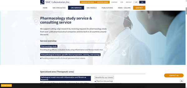
Reference: SMC Laboratories, Inc. official website (https://www.smccro-lab.com/)
SMC Laboratories, Inc.
SMC Laboratories is a specialized non-clinical CRO focused on in vivo pharmacology and efficacy studies using disease-relevant animal models, particularly in fibrosis, inflammation, metabolic diseases, and oncology.
Areas of Expertise
Disease-Relevant Model Portfolio
SMC Laboratories offers models covering the liver, lung, kidney, intestine, and oncology. Its portfolio includes the proprietary STAM™ model for MASH, fibrosis, and hepatocellular carcinoma.
Study Design Based on Target Biology
Study plans are developed around the target biology, mechanism of action, disease stage, and development objective. Pharmacological endpoints can be combined with histopathology, biomarkers, and disease-specific readouts.
Support from Target Validation to Proof of Concept
With experience from more than 1,000 studies for clients in 30 countries, SMC Laboratories supports programs from target validation and candidate selection through in vivo proof-of-concept studies.
Safety StudiesComprehensive Safety Assessment for Preclinical Development
Reference: Charles River Laboratories official website (https://www.criver.com/)
Charles River Laboratories
Charles River provides non-clinical toxicology and safety assessment services for programs ranging from exploratory safety studies to IND-enabling development.
Areas of Expertise
General Toxicology Across Study Designs
Services include single- and repeat-dose toxicology, dose-range finding, and general toxicology studies across multiple species and administration routes.
Non-GLP and GLP Study Support
Charles River supports both non-GLP and GLP studies, allowing sponsors to progress from early safety characterization to studies intended for regulatory submissions.
Integrated IND-Enabling Safety Assessment
Toxicology studies can be integrated with toxicokinetics, clinical pathology, histopathology, and safety pharmacology to support interpretation and IND-enabling safety packages.
Pharmacokinetic (PK/PD) StudiesConnecting Drug Exposure with Pharmacological Response
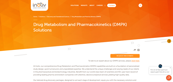
Reference: Inotiv official website (https://www.inotiv.com/)
Inotiv
Inotiv provides integrated PK/PD, DMPK, and bioanalytical services to characterize drug exposure and its relationship with pharmacological response.
Areas of Expertise
Pharmacokinetic Characterization
PK studies characterize exposure, half-life, clearance, and other pharmacokinetic parameters needed to understand how a candidate behaves in the selected model.
Exposure–Response Evaluation
Pharmacokinetic data can be combined with pharmacodynamic endpoints and bioanalysis to evaluate the relationship between drug exposure and pharmacological response.
Integrated DMPK and Development Support
Integrated DMPK, pharmacology, and safety information supports candidate comparison, dose selection, dosing-frequency optimization, and decisions about subsequent preclinical development.

 Japanese