Light-sheet fluorescence microscopy for 3D imaging
Light-sheet fluorescence microscopy (LSFM) illuminates a plane of the specimen and detects emitted light along an orthogonal axis.[S1] This page focuses on the imaging system: acquisition speed, illumination burden, field of view, resolution, data infrastructure, and contract-imaging deliverables. Clearing chemistry and whole-brain registration are addressed separately.
Why LSFM is used for large 3D specimens
LSFM restricts excitation largely to the observed plane, reducing out-of-focus illumination and phototoxicity while enabling faster volumetric acquisition than point-scanning approaches in many large-sample applications.[S1][S2] Cleared-tissue reviews pair LSFM with optical clearing and automated analysis,[S3] and the CUBIC work provides a whole-brain implementation example.[S4] The tradeoff is optical: increasing numerical aperture can improve resolution while narrowing the field of view, so no LSFM configuration is optimal for every specimen scale.[S2]
Three implementation constraints
Capital and operating requirements
An internal LSFM program requires an optical configuration matched to the specimen and enough storage and processing capacity for the resulting volume data.[S2][S5] Budget separately for sample chambers, optics, lasers and cameras, calibration and service, environmental control, acquisition workstations, storage, backup, and image-processing compute. Request a total-cost model based on expected sample volume and retention period. No equipment price is stated here because the research for this page did not verify a current US price.
Specimen and optical compatibility
The imager must accept the cleared specimen as delivered. Confirm refractive-index range, immersion medium, sample dimensions, mounting geometry, working distance, objective correction, excitation wavelengths, emission filters, and multiview capability before clearing begins. Run a pilot to define acceptable transparency, label penetration, background, striping, shadowing, stitching error, and usable imaging depth. Clearing chemistry itself belongs in the tissue-clearing protocol, not in the microscope specification.
Data handling and computation
A single LSFM experiment can generate data ranging from hundreds of gigabytes to petabytes, and published processing work identifies input-output throughput and memory as major constraints.[S5] Before acquisition, estimate raw, intermediate, and final volume; define file format and metadata; test transfer speed; set retention and backup periods; and identify who will perform stitching, deconvolution, registration, segmentation, and quality control. Compute architecture should follow the validated pipeline rather than assuming that a GPU alone solves the bottleneck.
When to use a contract imaging service
End-to-end scope
If clearing, labeling, acquisition, and analysis are outsourced together, place each stage under one statement of work; if they are divided, document every handoff. Decide where custody transfers occur, who owns method development, which intermediate artifacts are delivered, how failed samples are handled, and whether the raw images and processing parameters are included. Do not assume a shorter lead time; compare a documented schedule that includes pilot work, reruns, quality review, and data transfer.
Method-to-instrument fit
Ask each laboratory to demonstrate compatibility between its clearing protocol, sample holder, immersion medium, objective, field of view, and analysis pipeline. The useful evidence is a pilot on the relevant organ and label with predefined acceptance criteria, not a list of protocol names. Require representative raw tiles as well as rendered images so striping, shadowing, saturation, stitching, and depth-dependent signal loss can be evaluated.
Quantitative deliverables
Automated analysis is feasible but must be specified and validated for the assay. ClearMap combined immunostaining, LSFM, automated mapping, and activity analysis in a published whole-brain pipeline.[S6] For a contracted endpoint, request the algorithm version, training or thresholding procedure, ground-truth set, false-positive and false-negative estimates, region masks, exclusion rules, per-sample results, and enough intermediate output to reproduce the reported number.
Selection checklist
Select the optical configuration from specimen size, target resolution, label brightness, spectral channels, and required throughput. Then compare proposals on a relevant pilot, calibration and quality controls, data volume, secure transfer, retention, raw-data access, analysis validation, rerun policy, and schedule. LSFM can reduce illumination burden and accelerate volumetric acquisition in suitable designs,[S2] but it is one component of an assay and does not by itself make the biological conclusion more reliable or more predictive of a human outcome.
Whole-brain imaging with single-cell resolution using chemical cocktails and computational analysis (CUBIC). https://europepmc.org/article/MED/24746791 (accessed 2026-09-03)
3 Recommended Contract Research Organizations
for Non-Clinical Studies
— by Target goal and Expertise
In non-clinical development, the choice of contract research organization shapes the quality of the data and the time it takes to reach the next decision. Below, three CROs are introduced by the type of study they support: pharmacology (efficacy) studies, safety studies, and pharmacokinetic (PK/PD) studies. Each summary describes the services the company offers so that you can match a provider to your target and development objective.
Pharmacology (Efficacy) StudiesDisease-Relevant Models for Translational Drug Evaluation
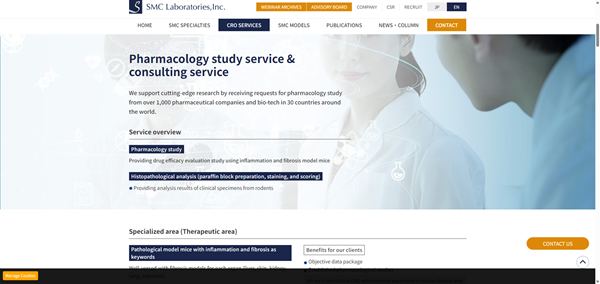
Reference: SMC Laboratories, Inc. official website (https://www.smccro-lab.com/)
SMC Laboratories, Inc.
SMC Laboratories is a specialized non-clinical CRO focused on in vivo pharmacology and efficacy studies using disease-relevant animal models, particularly in fibrosis, inflammation, metabolic diseases, and oncology.
Areas of Expertise
Disease-Relevant Model Portfolio
SMC Laboratories offers models covering the liver, lung, kidney, intestine, and oncology. Its portfolio includes the proprietary STAM™ model for MASH, fibrosis, and hepatocellular carcinoma.
Study Design Based on Target Biology
Study plans are developed around the target biology, mechanism of action, disease stage, and development objective. Pharmacological endpoints can be combined with histopathology, biomarkers, and disease-specific readouts.
Support from Target Validation to Proof of Concept
With experience from more than 1,000 studies for clients in 30 countries, SMC Laboratories supports programs from target validation and candidate selection through in vivo proof-of-concept studies.
Safety StudiesComprehensive Safety Assessment for Preclinical Development
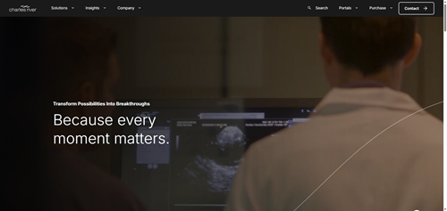
Reference: Charles River Laboratories official website (https://www.criver.com/)
Charles River Laboratories
Charles River provides non-clinical toxicology and safety assessment services for programs ranging from exploratory safety studies to IND-enabling development.
Areas of Expertise
General Toxicology Across Study Designs
Services include single- and repeat-dose toxicology, dose-range finding, and general toxicology studies across multiple species and administration routes.
Non-GLP and GLP Study Support
Charles River supports both non-GLP and GLP studies, allowing sponsors to progress from early safety characterization to studies intended for regulatory submissions.
Integrated IND-Enabling Safety Assessment
Toxicology studies can be integrated with toxicokinetics, clinical pathology, histopathology, and safety pharmacology to support interpretation and IND-enabling safety packages.
Pharmacokinetic (PK/PD) StudiesConnecting Drug Exposure with Pharmacological Response
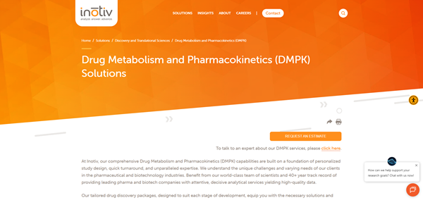
Reference: Inotiv official website (https://www.inotiv.com/)
Inotiv
Inotiv provides integrated PK/PD, DMPK, and bioanalytical services to characterize drug exposure and its relationship with pharmacological response.
Areas of Expertise
Pharmacokinetic Characterization
PK studies characterize exposure, half-life, clearance, and other pharmacokinetic parameters needed to understand how a candidate behaves in the selected model.
Exposure–Response Evaluation
Pharmacokinetic data can be combined with pharmacodynamic endpoints and bioanalysis to evaluate the relationship between drug exposure and pharmacological response.
Integrated DMPK and Development Support
Integrated DMPK, pharmacology, and safety information supports candidate comparison, dose selection, dosing-frequency optimization, and decisions about subsequent preclinical development.

 Japanese